Steps of Laparoscopic Endometriosis Surgery
Here are the sequential actions involved in performing laparoscopic surgery for endometriosis.
Laparoscopic endometriosis surgery, also known as minimally invasive surgery or keyhole surgery, is a procedure used to diagnose and treat endometriosis. Here are the general steps involved in the surgery:
Step 1: Anesthesia
There are generally two types of anesthesia used in laparoscopic endometriosis surgery:
- General Anesthesia: This is the most common type of anesthesia used in laparoscopic procedures. It involves administering medications to induce a state of unconsciousness, where the patient is completely unaware and does not feel any pain during the surgery. General anesthesia is typically administered through intravenous (IV) medications and inhalation agents.
- Regional Anesthesia: In some cases, regional anesthesia may be used as an alternative or in combination with general anesthesia. Regional anesthesia involves numbing a specific region of the body, such as the lower abdomen and pelvic area, using local anesthetics. This can be achieved through techniques such as epidural anesthesia or spinal anesthesia. Regional anesthesia can provide pain relief during and after the surgery, and it may also help reduce the need for high doses of general anesthesia.
Step 2: Patient Positioning
The patient is positioned on the operating table in a way that allows the surgeon to access the pelvic region. The legs are usually elevated and placed in stirrups to provide optimal exposure.
Step 3: Creation of Pneumoperitoneum
A small incision is made near the navel, and a hollow needle or Veress needle is inserted into the abdominal cavity. Carbon dioxide gas is then pumped into the abdomen, creating a pneumoperitoneum.
The primary goal of pneumoperitoneum is to create a working space within the abdomen, separating the abdominal wall from the organs. This inflated space provides the surgeon with the necessary room to maneuver instruments and perform the procedure effectively.
Carbon dioxide (CO2) gas is the preferred gas for creating pneumoperitoneum. It is non-flammable, readily absorbed by the body, and has a minimal impact on organ function. CO2 is usually delivered into the abdominal cavity in a controlled manner to maintain a stable pressure.
Step 4: Placement of Trocars
Trocars are specialized instruments with a valve that allow entry into the abdominal cavity while maintaining the pneumoperitoneum. Several small incisions, usually ranging from 0.5 to 1 centimeter in size, are made in the abdominal wall, and trocars are inserted through these incisions. These trocars act as ports for the laparoscope and other surgical instruments.
Step 5: Visualization with a Laparoscope
A laparoscope, a long, thin tube with a camera and light source at the tip, is inserted through one of the trocars. The laparoscope transmits real-time images of the pelvic organs to a monitor, allowing the surgeon to visualize the surgical site.
Step 6: Exploration and Identification
The surgeon examines the pelvic region, looking for signs of endometriosis. Various instruments, such as dissectors, forceps, and scissors, may be used to gently manipulate and move organs to get a better view.
Step 7: Excision or Ablation of Endometriotic Lesions
During laparoscopic endometriosis surgery, one of the main objectives is to treat the endometriotic lesions found in the pelvic region. There are two common methods used to address these lesions: excision and ablation. Here’s an explanation of each approach:
- Excision: Excision involves physically cutting out the endometriotic lesions from the affected tissues. The surgeon uses specialized instruments, such as scissors, electrocautery devices, or lasers, to carefully remove the lesions while preserving the surrounding healthy tissue. Excision is often preferred for deeper or larger endometriotic lesions that have invaded the surrounding structures or organs. It aims to completely eliminate the lesions and can provide a more thorough treatment.
- Ablation: Ablation refers to the destruction or removal of endometriotic lesions using energy-based techniques. The surgeon applies heat, electrical energy, laser energy, or other modalities to destroy the lesions. This process can be done using various instruments, such as electrodes, lasers, or radiofrequency devices. Ablation is often used for superficial or smaller endometriotic lesions that have not deeply infiltrated the surrounding tissues. While ablation can be effective in removing the lesions, there is a higher chance of residual disease compared to excision.
The choice between excision and ablation depends on factors such as the location, size, and depth of the lesions, as well as the surgeon’s expertise and the extent of the disease. In some cases, a combination of both techniques may be utilized during the same procedure to address different types or locations of endometriotic lesions.
Step 8: Hemostasis
Any bleeding from the excised or ablated areas is carefully controlled. The surgeon may use electrocautery, laser, or other techniques to achieve hemostasis and prevent excessive bleeding.
Step 9: Closure and Removal of Instruments
Once the surgery is complete, the surgeon removes the instruments and trocars from the abdominal cavity. The small incisions in the abdominal wall are closed with sutures or surgical glue.
Step 10: Recovery
The patient is transferred to the recovery room, where they are monitored until they wake up from anesthesia. Pain medications and instructions for postoperative care are provided. The patient usually goes home on the same day or within a day or two, depending on the complexity of the surgery.
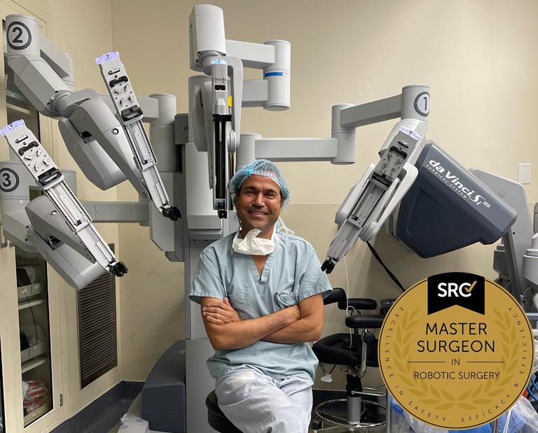
Pankaj Singhal, MD, MS, MHCM
Master Surgeon in Robotic Surgery
Dr. Pankaj Singhal, a globally recognized endometriosis surgeon, possesses over 25 years of expertise in laparoscopic excision surgery, enabling him to tackle even the most challenging endometriosis cases with confidence. Dr. Pankaj treats patients with diverse endometriosis-related conditions, ranging from ovarian endometriomas to severe deep infiltrating endometriosis that affects the bowels and other organs.
Dr. Pankaj prioritizes minimally invasive surgery and provides comprehensive personal care. Additionally, he is the owner and founder of New York Gynecology and Endometriosis (NYGE), and has dedicated his life to advocating for, respecting, and treating women suffering from this little-known disease. He is one of the few surgeons in the entire United States who have completed over 5,718 robot-assisted gynecologic surgeries.

We Accept Most Major Insurance Plans
Convenient Billing Options for Comprehensive Coverage.
Surgeries are typically covered by health insurance. However, the extent of coverage can vary depending on the specific insurance plan and policy. Some insurance plans may cover a broad range of surgical procedures, including both elective and necessary surgeries, while others may have limitations or exclusions for certain procedures.
In some cases, certain insurance plans or programs may fully cover the cost of surgery, leaving the patient with no financial responsibility.
Request an Appointment with
New York Gynecology Endometriosis
"*" indicates required fields

