
Adenomyosis
Adenomyosis is a condition in which the lining tissue of the uterus grows into the muscle wall of the uterus, causing pain and heavy menstrual bleeding.
Adenomyosis can coexist with other gynecological conditions such as endometriosis, fibroids, and pelvic inflammatory disease.
It is a common condition, affecting an estimated 1 in 10 women of reproductive age, and it is most commonly diagnosed in women between the ages of 40 and 50.
Differences Between Adenomyosis and Endometriosis
Adenomyosis and endometriosis are two distinct but related gynecological conditions.
Adenomyosis is a condition in which the tissue that is similar to the lining of the uterus (endometrium) grows into the muscle wall of the uterus, causing the uterus to become enlarged and painful during menstruation.
Endometriosis, on the other hand, is a condition in which the tissue that is similar to the lining of the uterus (endometrial tissue) grows outside of the uterus and on other organs such as the ovaries, fallopian tubes, and pelvic lining, leading to pain and infertility.
While both conditions involve the presence of endometrial tissue outside of its normal location, adenomyosis is limited to the uterus, whereas endometriosis can occur in other areas of the pelvis and even outside of the pelvis. Additionally, the symptoms of the two conditions can overlap, but they are not identical.
Causes of Adenomyosis
Over the past decade, numerous studies have identified multiple factors that could potentially cause adenomyosis, including sex hormone receptors, inflammatory molecules, enzymes in the extracellular matrix, growth factors, and neuroangiogenic factors.
There are several theories surrounding the development of adenomyosis, including:
- Invasive growth of endometrial tissue: Some researchers believe that adenomyosis develops when endometrial tissue grows into the muscle layer of the uterus, causing it to become thick and swollen.
- Hormonal imbalances: It has been suggested that hormonal imbalances, particularly increased levels of estrogen, may contribute to the development of adenomyosis.
- Inflammation: Inflammation of the uterus may also play a role in the development of adenomyosis, as it can lead to the migration of endometrial tissue into the muscle layer of the uterus.
- Prior uterine surgery: Women who have had prior uterine surgery, such as a cesarean delivery or fibroid removal, may be more likely to develop adenomyosis.
- Genetic factors: There may also be a genetic component to the development of adenomyosis, as it appears to run in some families.
Factors That Increase the Risk of Adenomyosis
There are several factors that have been associated with an increased risk of developing adenomyosis, including:
- Age: Adenomyosis is most commonly diagnosed in women between the ages of 40 and 50, suggesting that hormonal changes associated with aging may increase the risk.
- Multiple pregnancies: Women who have had multiple pregnancies may have an increased risk of developing adenomyosis.
Diagnosis of Adenomyosis
Diagnosing adenomyosis typically involves a combination of medical history, physical examination, and imaging studies. In some cases, a biopsy of the uterine tissue may be necessary to confirm the diagnosis. It is important to note that the definitive diagnosis of adenomyosis can only be made through a histological examination of the uterus.
Imaging techniques used to diagnose adenomyosis include ultrasound, magnetic resonance imaging (MRI), and hysteroscopy.
- Ultrasound is often the initial imaging technique used to evaluate for adenomyosis, as it is non-invasive, widely available, and relatively inexpensive. Transvaginal ultrasound, in particular, can provide high-resolution images of the uterus and can be used to identify the characteristic features of adenomyosis, such as thickening of the uterine wall, the presence of cysts, and asymmetry of the uterus.
- MRI is another imaging technique that can provide detailed images of the uterus and is particularly useful in cases where adenomyosis is suspected but not clearly visible on ultrasound. MRI can provide better visualization of the deep myometrial involvement and extent of adenomyosis lesions.
- Hysteroscopy involves inserting a thin, flexible tube with a camera through the cervix and into the uterus to directly visualize the uterine lining and muscular wall. It can be used to diagnose adenomyosis, as well as other conditions such as fibroids or polyps, and can also be used for treatment purposes.
Classifications of Adenomyosis
Adenomyosis can be classified based on the distribution and extent of the endometrial tissue within the uterine wall:
- Focal adenomyosis: This type of adenomyosis is characterized by discrete, well-defined lesions or nodules within the uterine wall.
- Diffuse adenomyosis: This type of adenomyosis is characterized by a uniform, widespread involvement of the uterine wall.
- Adenomyoma: This type of adenomyosis is a variant where the endometrial tissue forms a distinct mass within the uterine wall, and can be mistaken for a fibroid.
- Cystic adenomyosis: This type of adenomyosis is characterized by the formation of cysts within the uterine wall, which may be filled with blood or other fluids.
- Adenomyosis externa: This type of adenomyosis refers to the presence of endometrial tissue outside of the uterus, often involving nearby structures such as the ovaries or fallopian tubes.
Accurate identification of the specific type of adenomyosis is crucial, as the choice of treatment approach will vary depending on the type of adenomyosis. For instance, in cases of focal adenomyosis and adenomyoma, appropriate surgical management can often avoid the need for a hysterectomy.
Can Adenomyosis Impact Pregnancy and Fertility?
Adenomyosis can affect pregnancy in several ways. It is associated with an increased risk of infertility and miscarriage, as well as complications during pregnancy such as preterm labor, abnormal fetal position, and postpartum hemorrhage. The presence of adenomyosis can also increase the likelihood of requiring a cesarean section delivery. Additionally, adenomyosis can cause pain and discomfort during pregnancy, which may require medical management.
Adenomyosis is associated with infertility, although the exact mechanisms are not well understood. It is thought that the abnormal uterine environment caused by adenomyosis, including inflammation and changes in the endometrial lining, can impair implantation of a fertilized egg and lead to difficulty getting pregnant. Adenomyosis may also interfere with the normal function of the fallopian tubes or hinder the movement of sperm.
A study presented at the Japan Society of OB/GYN reported that women with adenomyosis had a higher risk of miscarriage (50%), pre-term birth (24.4%), and fetal retardation (nearly 12%) compared to women without adenomyosis.
However, with appropriate monitoring and management, many women with adenomyosis are able to have successful pregnancies.
In some cases, assisted reproductive technologies such as in vitro fertilization (IVF) may be recommended to improve the chances of pregnancy.
- Differences Between Adenomyosis and Endometriosis
- Causes of Adenomyosis
- Factors that Increase the Risk of Adenomyosis
- Diagnosis of Adenomyosis
- Classifications of Adenomyosis
- Adenomyosis: Pregnancy and Fertility
- Surgical Methods for Adenomyosis Treatment
- - Laparoscopic Deep Excisional Adenomyosis Surgery (LEAS)
- - Robotic-Assisted Laparoscopy
- - Laparotomy
- - Osada Procedure
Surgical Methods for Adenomyosis Treatment
There are several surgical methods for treating adenomyosis, including Laparoscopic deep excisional adenomyosis surgery (LEAS), robotic-assisted laparoscopy, laparotomy, and Osada procedure. The specific method used depends on the severity and location of the adenomyosis tissue, as well as the patient’s overall health and preferences. In general, the goal of surgery for adenomyosis is to remove the affected tissue while preserving healthy tissue and organs as much as possible.
Laparoscopic Deep Excisional Adenomyosis Surgery (LEAS)
Laparoscopic deep excisional adenomyosis surgery (LEAS) is a minimally invasive surgical procedure used to treat adenomyosis.
During the surgery, a laparoscope is used to visualize the inside of the uterus, and the adenomyotic tissue is excised from the walls of the uterus. LEAS is a specialized surgical technique that involves removing the adenomyotic tissue as deeply as possible, often using specialized instruments and techniques to ensure complete removal of the affected tissue.
This surgery is typically reserved for women with more severe or extensive adenomyosis, who have not responded to other treatments, and who wish to preserve their fertility.
Robotic-Assisted Laparoscopy
Robotic-assisted laparoscopy is a minimally invasive surgical technique that can be used to treat adenomyosis.
During the procedure, a surgeon controls robotic arms equipped with surgical instruments to perform the surgery through small incisions in the abdomen. This approach allows for more precise movements and enhanced visualization compared to traditional laparoscopy. Robotic-assisted laparoscopy may be a suitable option for some women with adenomyosis, particularly those with more complex cases, but it is not always necessary or appropriate for every patient.
The choice of surgical approach will depend on individual circumstances, the extent and severity of the adenomyosis, and other factors that need to be taken into account when deciding on the best treatment option.
Laparotomy
Laparotomy is a more invasive surgical procedure that can be used to treat adenomyosis. During a laparotomy, a larger incision is made in the abdomen to allow the surgeon to directly access the uterus and other reproductive organs. This approach may be necessary in cases where the adenomyosis is more extensive or severe, or if other treatments have not been effective.
However, laparotomy is generally reserved for more complex cases, and is associated with a longer recovery time and a higher risk of complications compared to minimally invasive approaches such as laparoscopic surgery.
Osada Procedure
The Osada procedure is a surgical technique used to treat adenomyosis. It involves removing the adenomyotic tissue from the uterus while preserving the uterus itself.
During the procedure, the uterus is separated from surrounding tissues, and the adenomyotic tissue is excised using a combination of surgical instruments and a laser. The uterine wall is then reconstructed and sutured closed. The Osada procedure is a specialized surgical technique that is typically reserved for women with adenomyosis who want to preserve their fertility, as it aims to remove the affected tissue while leaving the uterus intact.
However, it is important to note that not all women with adenomyosis will be candidates for this procedure, and the choice of surgical approach will depend on individual circumstances and factors such as the extent and severity of the adenomyosis, overall health, and reproductive goals.
Adenomyosis Non-surgical Treatment
Although non-surgical treatments like NSAIDs, progestin-only birth control pills, and GnRH analogs can alleviate some symptoms of adenomyosis, they are usually temporary and do not provide a complete cure for the disease. The most effective approach to treating adenomyosis involves undergoing laparoscopic deep excision surgery and a comprehensive multidisciplinary treatment plan.
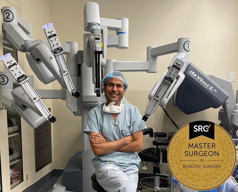
Pankaj Singhal, MD, MS, MHCM
Master Surgeon in Robotic Surgery
Dr. Pankaj Singhal, a globally recognized endometriosis surgeon, possesses over 25 years of expertise in laparoscopic excision surgery, enabling him to tackle even the most challenging endometriosis cases with confidence. Dr. Pankaj treats patients with diverse endometriosis-related conditions, ranging from ovarian endometriomas to severe deep infiltrating endometriosis that affects the bowels and other organs.
Dr. Pankaj prioritizes minimally invasive surgery and provides comprehensive personal care. Additionally, he is the owner and founder of New York Gynecology and Endometriosis (NYGE), and has dedicated his life to advocating for, respecting, and treating women suffering from this little-known disease. He is one of the few surgeons in the entire United States who have completed over 5,718 robot-assisted gynecologic surgeries.
We Accept Most Major Insurance Plans
Convenient Billing Options for Comprehensive Coverage.
Surgeries are typically covered by health insurance. However, the extent of coverage can vary depending on the specific insurance plan and policy. Some insurance plans may cover a broad range of surgical procedures, including both elective and necessary surgeries, while others may have limitations or exclusions for certain procedures.
In some cases, certain insurance plans or programs may fully cover the cost of surgery, leaving the patient with no financial responsibility.
Request an Appointment with
New York Gynecology Endometriosis
"*" indicates required fields