Comprehensive List of All Endometriosis Cases That We Treat
Most Complex Robotic Endometriosis Cases
- Bowel, bladder, ureters, nerves are managed by us.
- Endometriosis is not cancer but grows like one.
- Being a cancer surgeon allows us to resect endometriosis with unparalleled precision.
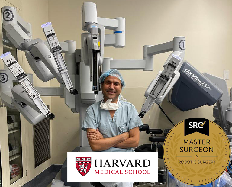
- Performed more than 10,000 robotic gynecologic, endometriosis and cancer surgeries.
- Known for taking on the most challenging surgery cases that other doctors/centers turn away.
Endometriosis of Uterus
The uterus is a pear-shaped organ in the female reproductive system responsible for housing and nourishing a developing fetus during pregnancy.
Deep endometriosis of the uterus involves the infiltration of endometrial tissue into the deeper layers of the uterine muscle, potentially causing pain, scarring, and distortion of the uterine structure. Superficial endometriosis of the uterus refers to the presence of endometrial tissue on the surface of the uterine lining without penetrating deeply into the muscle layer.
Adenomyosis of the uterus is a separate condition where endometrial tissue grows into the muscular wall of the uterus, leading to thickening of the uterine wall, painful menstruation, and sometimes heavy bleeding. While all these conditions involve endometrial tissue, they differ in terms of the depth of tissue infiltration and specific location within or around the uterus, contributing to variations in symptoms and potential complications.
Endometriosis of Ovary
The ovary is an essential reproductive organ in females responsible for producing eggs and releasing hormones. Deep endometriosis of the ovary involves the infiltration of endometrial tissue into the deeper layers of the ovary, potentially forming cysts called endometriomas and leading to scarring and distortion of ovarian tissue. Superficial endometriosis of the ovary refers to the presence of endometrial tissue on the surface of the ovary without infiltration into its deeper layers.
Both types of endometriosis of the ovary can cause pelvic pain and may impact fertility, but deep endometriosis tends to be associated with more severe symptoms and potential complications due to the involvement of deeper ovarian structures.
Endometriosis of Fallopian Tube
The fallopian tube is a slender, tube-like structure connecting the ovaries to the uterus, serving as a passageway for eggs released by the ovaries to travel to the uterus.
Deep endometriosis of the fallopian tube involves the infiltration of endometrial tissue into the deeper layers of the tube, potentially causing inflammation, scarring, and blockages that may impair fertility. Superficial endometriosis of the fallopian tube refers to the presence of endometrial tissue on the outer surface of the tube without infiltration into its deeper layers.
Both types of endometriosis of the fallopian tube can lead to symptoms such as pelvic pain and may impact fertility, though the severity and specific implications can vary between individuals.
Endometriosis of Pelvic Peritoneum
The pelvic peritoneum refers to the membrane lining the pelvic cavity, providing support and protection to pelvic organs. The anterior cul-de-sac is the space between the bladder and uterus in females. The posterior cul-de-sac is the space between the uterus and rectum. The pelvic sidewall consists of the muscles and connective tissues along the sides of the pelvic cavity, contributing to pelvic support and stability. The pelvic brim is the boundary between the pelvic cavity and the abdominal cavity, demarcating the entrance to the pelvis. The uterosacral ligaments are fibrous bands that attach the uterus to the sacrum, providing structural support to the uterus and helping maintain its position within the pelvis.
Endometriosis of Rectovaginal Septum and Vagina
The rectovaginal septum is the anatomical structure separating the rectum from the vagina, while the vagina is a muscular canal connecting the external genitalia to the cervix, forming part of the female reproductive system.
Endometriosis of the rectovaginal septum and vagina refers to the presence of endometrial-like tissue growth in these respective anatomical regions, potentially causing symptoms such as pelvic pain, painful intercourse, and discomfort.
Endometriosis of Intestine
The intestine is a vital component of the digestive system, comprising the small intestine, where nutrient absorption primarily occurs, and the large intestine, which includes the sigmoid colon, cecum, appendix, and various other segments such as the ascending, transverse, and descending colon. The sigmoid colon, forming the final part of the large intestine before the rectum, stores feces before elimination, while the rectum serves as the final section of the large intestine where feces are stored before expulsion. The cecum, located at the beginning of the large intestine, aids in fluid and salt absorption. The appendix, a small extension attached to the cecum, has debated functions potentially related to the immune system.
Overall, these structures work synergistically to process food, absorb nutrients, and facilitate waste elimination in the human body’s digestive process.
Endometriosis of Cutaneous Scar
Endometriosis of a cutaneous scar occurs when endometrial tissue implants and grows within a surgical or traumatic scar on the skin, resulting in pain, swelling, and the formation of nodules or cysts at the scar site. The prevalence of endometriosis occurring within cutaneous scars is relatively low, estimated to affect around 1% to 2% of individuals with endometriosis.
Endometriosis of Bladder and Ureters
The bladder is a hollow organ located in the pelvis responsible for storing urine before it is expelled from the body, while the ureters are narrow tubes that carry urine from the kidneys to the bladder.
Deep endometriosis of the bladder or ureters involves the infiltration of endometrial tissue into the deeper layers of these organs, potentially causing inflammation, scarring, and obstruction of urine flow, leading to symptoms such as pelvic pain, urinary urgency, and recurrent urinary tract infections. Superficial endometriosis of the bladder or ureters, however, refers to the presence of endometrial tissue on the surface of these organs without penetrating deeply into their layers.
Both types of endometriosis of the bladder or ureters can result in significant discomfort and may require a combination of medical and surgical interventions for management.
Endometriosis of Cardiothoracic Space
Endometriosis of the cardiothoracic space refers to the presence of endometrial-like tissue growth within the chest cavity, potentially affecting structures such as the pleura, lung and mediastinal spaces.
Pleural endometriosis involves the infiltration of endometrial tissue into the lining of the lungs and chest cavity, leading to symptoms such as chest pain and shortness of breath. Lung endometriosis occurs when endometrial-like implants are found within the lung tissue, sometimes causing symptoms like coughing up blood or chest discomfort. Mediastinal endometriosis involves the growth of endometrial-like tissue within the mediastinum, the space in the middle of the chest containing the heart, esophagus, and other vital structures.
Deep endometriosis of the diaphragm refers to the infiltration of endometrial tissue into the muscular or connective tissue layers of the diaphragm, potentially causing referred pain and respiratory symptoms. Superficial endometriosis of the diaphragm, however, describes the presence of endometrial tissue on the surface of the diaphragm without penetrating deeply into its layers.
Endometriosis of Abdomen
Abdominal endometriosis refers to the presence of endometrial tissue outside the uterus within the abdomen, often causing pain and infertility.
The umbilicus, commonly known as the belly button, is a central point on the abdomen where the umbilical cord was attached during fetal development. The inguinal canal is a passage in the lower abdomen through which structures like the spermatic cord in males or the round ligament of the uterus in females pass.
Extra-pelvic abdominal peritoneum refers to the peritoneal lining that extends beyond the pelvic cavity into the abdomen, serving as a protective membrane for abdominal organs. The anterior abdominal wall is the front portion of the abdomen, consisting of layers of muscle and fascia that provide support and protection for the abdominal organs.
Endometriosis of Pelvic Nerves
Endometriosis of the pelvic nerves involves the infiltration of endometrial tissue into the nerves within the pelvic region, potentially leading to neuropathic pain and dysfunction.
Sacral splanchnic nerves are responsible for transmitting sensory and motor signals between the pelvic organs and the sacral spinal cord, and endometriosis in these nerves can cause pelvic pain and disruptions in organ function. Sacral nerve roots originate from the sacral region of the spinal cord and can be affected by endometriosis, contributing to pelvic pain and discomfort. Endometriosis involving the obturator nerve, which innervates the inner thigh and pelvic region, can result in pain during movement and intercourse.
Endometriosis affecting the sciatic nerve, the largest nerve in the body, can lead to radiating pain, numbness, and weakness in the lower back, buttocks, and legs. The pudendal nerve, responsible for sensation in the genital and perineal area, can be impacted by endometriosis, causing pelvic pain and sexual dysfunction. Endometriosis involving the femoral nerve, which supplies sensation to the thigh and leg, may result in pain and weakness in these regions, potentially affecting mobility and quality of life.
Pankaj Singhal, MD, MS, MHCM, FACOG
Dr. Singhal previously served as the Chairman of the Department of Obstetrics & Gynecology at Good Samaritan and was the System Chairman for Obstetrics, Gynecology, and Women’s Health for Catholic Health Services. He obtained his medical degree from Madras Medical College in India and holds a master’s degree in healthcare management from Harvard University.
Dr. Singhal is an accredited Surgeon of Excellence by the Surgical Review Corporation (SRC) and has received the Center of Excellence designation in Robotic Surgery and Minimally Invasive Gynecology from Good Samaritan.
Dr. Singhal’s special interests and expertise are in advanced reproductive technologies, laparoscopic and minimally invasive surgery, gynecology, and endometriosis. He consults with and provides assistance to clients from all over the world in various areas of gynecology surgery. He is recognized for taking on the most challenging surgery and endometriosis cases that other doctors/centers decline. His existing and past clients have been his biggest supporters, referring him to their friends and family. Dr. Singhal’s ongoing new client base is primarily made up of referrals from prior clients.
As a leader in robotic and minimally invasive surgery, Dr. Singhal has established the New York Gynecology Endometriosis (NYGE) center, which emphasizes a thorough and cautious treatment approach that addresses all of women’s endometriosis-related requirements.

Dr. Singhal's Research Publications Across Various Platforms
- Singhal, P., & Lele, S. (2006). Intraperitoneal chemotherapy for ovarian cancer: where are we now?. Journal of the National Comprehensive Cancer Network : JNCCN, 4(9), 941–946. https://doi.org/10.6004/jnccn.2006.0077
- Singhal, P., Odunsi, K., Rodabaugh, K., Driscoll, D., & Lele, S. (2006). Primary fallopian tube carcinoma: a retrospective clinicopathologic study. European journal of gynaecological oncology, 27(1), 16–18.
- Hylander, B., Repasky, E., Shrikant, P., Intengan, M., Beck, A., Driscoll, D., Singhal, P., Lele, S., & Odunsi, K. (2006). Expression of Wilms tumor gene (WT1) in epithelial ovarian cancer. Gynecologic oncology, 101(1), 12–17. https://doi.org/10.1016/j.ygyno.2005.09.052
- McCloskey, S. A., Tchabo, N. E., Malhotra, H. K., Odunsi, K., Rodabaugh, K., Singhal, P., Lele, S., & Jaggernauth, W. (2010). Adjuvant vaginal brachytherapy alone for high risk localized endometrial cancer as defined by the three major randomized trials of adjuvant pelvic radiation. Gynecologic oncology, 116(3), 404–407. https://doi.org/10.1016/j.ygyno.2009.06.027
- Singhal, P., Spiegel, G., Driscoll, D., Odunsi, K., Lele, S., & Rodabaugh, K. J. (2005). Cyclooxygenase 2 expression in serous tumors of the ovary. International journal of gynecological pathology : official journal of the International Society of Gynecological Pathologists, 24(1), 62–66.
- McEachron, J., Zhou, N., Spencer, C., Shanahan, L., Chatterton, C., Singhal, P., & Lee, Y. C. (2020). Evaluation of the optimal sequence of adjuvant chemotherapy and radiation therapy in the treatment of advanced endometrial cancer. Journal of gynecologic oncology, 31(6), e90. https://doi.org/10.3802/jgo.2020.31.e90
- Singhal, P., Tchabo, N. E., & Odunsi, K. (2007). Immunologic markers of cancer progression and prognosis. Expert opinion on medical diagnostics, 1(4), 439–450. https://doi.org/10.1517/17530059.1.4.439
- Shrikant, P. A., Rao, R., Li, Q., Kesterson, J., Eppolito, C., Mischo, A., & Singhal, P. (2010). Regulating functional cell fates in CD8 T cells. Immunologic research, 46(1-3), 12–22. https://doi.org/10.1007/s12026-009-8130-9
- Eddib, A., Danakas, A., Hughes, S., Erk, M., Michalik, C., Narayanan, M. S., Krovi, V., & Singhal, P. (2014). Influence of Morbid Obesity on Surgical Outcomes in Robotic-Assisted Gynecologic Surgery. Journal of gynecologic surgery, 30(2), 81–86. https://doi.org/10.1089/gyn.2012.0142
- McEachron, J., Zhou, N., Spencer, C., Chatterton, C., Shanahan, L., Katz, J., Naegele, S., Singhal, P., & Lee, Y. C. (2021). Adjuvant chemoradiation associated with improved outcomes in patients with microsatellite instability-high advanced endometrial carcinoma. International journal of gynecological cancer : official journal of the International Gynecological Cancer Society, 31(2), 203–208. https://doi.org/10.1136/ijgc-2020-001709
- McEachron, J., Heyman, T., Shanahan, L., Tran, V., Friedman, M., Gorelick, C., Economos, K., Singhal, P., Lee, Y. C., & Kanis, M. J. (2020). Multimodality adjuvant therapy and survival outcomes in stage I-IV uterine carcinosarcoma. International journal of gynecological cancer : official journal of the International Gynecological Cancer Society, 30(7), 1012–1017. https://doi.org/10.1136/ijgc-2020-001315
- Eisner, I. S., Wadhwa, R. K., Downing, K. T., & Singhal, P. (2019). Prevention and management of bowel injury during gynecologic laparoscopy: an update. Current opinion in obstetrics & gynecology, 31(4), 245–250. https://doi.org/10.1097/GCO.0000000000000552
- Jun, S. K., Sathia Narayanan, M., Singhal, P., Garimella, S., & Krovi, V. (2013). Evaluation of robotic minimally invasive surgical skills using motion studies. Journal of robotic surgery, 7(3), 241–249. https://doi.org/10.1007/s11701-013-0419-y
- Eddib, A., Jain, N., Aalto, M., Hughes, S., Eswar, A., Erk, M., Michalik, C., Krovi, V., & Singhal, P. (2013). An analysis of the impact of previous laparoscopic hysterectomy experience on the learning curve for robotic hysterectomy. Journal of robotic surgery, 7(3), 295–299. https://doi.org/10.1007/s11701-012-0388-6
- Bolnick, A. D., Bolnick, J. M., Kohan-Ghadr, H. R., Kilburn, B. A., Pasalodos, O. J., Singhal, P., Dai, J., Diamond, M. P., Armant, D. R., & Drewlo, S. (2017). Enhancement of trophoblast differentiation and survival by low molecular weight heparin requires heparin-binding EGF-like growth factor. Human reproduction (Oxford, England), 32(6), 1218–1229. https://doi.org/10.1093/humrep/dex069
- Karabakhtsian, R., Heller, D. S., Singhal, P., & Sama, J. (2002). Malignant mixed mesodermal tumor in a young woman with polycystic ovary syndrome. A case report. The Journal of reproductive medicine, 47(11), 946–948.
- Castillo, M. H., Peoples, J. B., Machicao, C. N., & Singhal, P. (2001). The lateral island trapezius myocutaneous flap for circumferential reconstruction of hypopharynx and cervical esophagus. Digestive surgery, 18(2), 93–97. https://doi.org/10.1159/000050107
- S. Kumar, P. Singhal and V. N. Krovi, “Computer-Vision-Based Decision Support in Surgical Robotics,” in IEEE Design & Test, vol. 32, no. 5, pp. 89-97, Oct. 2015, doi: 10.1109/MDAT.2015.2465135. https://ieeexplore.ieee.org/document/7181689
- Kumar, Suren & Sathia narayanan, Madusudanan & Singhal, Pankaj & Corso, Jason & Krovi, Venkat. (2014). Surgical Tool Attributes from Monocular Video. 10.13140/2.1.2057.4081. https://www.researchgate.net/publication/264972097_Surgical_Tool_Attributes_from_Monocular_Video
- Eddib, A. , Hughes, S. , Aalto, M. , Eswar, A. , Erk, M. , Michalik, C. , Krovi, V. and Singhal, P. (2014) Impact of Age on Surgical Outcomes after Robot Assisted Laparoscopic Hysterectomies. Surgical Science, 5, 90-96. doi: 10.4236/ss.2014.53018.
- Chitgar, S., Ghomi, A., Danakas, G., Erk, M., Narayanan, M.S., Krovi, V., Sharma, S., and Singhal, P., “Safety and Efficacy of Robot-Assisted Gynecologic Surgery for High Risk and Advanced Stage Endometrial Cancer,” Journal of Minimally Invasive Gynecology , Vol. 20 , No. 6 , S82, Nov. 2013. doi:10.1016/j.jmig.2013.08.264.
We Accept Most Major Insurance Plans
Convenient Billing Options for Comprehensive Coverage.
Surgeries are typically covered by health insurance. However, the extent of coverage can vary depending on the specific insurance plan and policy. Some insurance plans may cover a broad range of surgical procedures, including both elective and necessary surgeries, while others may have limitations or exclusions for certain procedures.
In some cases, certain insurance plans or programs may fully cover the cost of surgery, leaving the patient with no financial responsibility.
Request an Appointment with
New York Gynecology Endometriosis
"*" indicates required fields
